
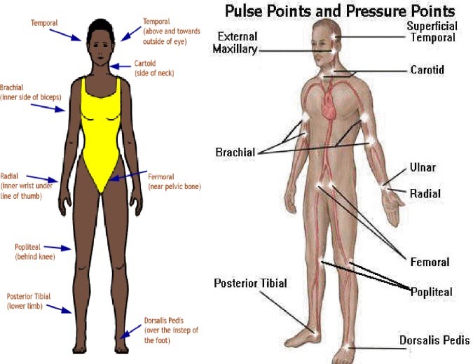
- Pulse is simply your cardiac performance that can be palpated at the neck (carotid), at the side of your head just above and lateral to the eye (temporal), at your chest specifically on the left side of the (apical), at the wrist (radial), at the inner aspect of the biceps (brachial), at the inguinal area (femoral), behind the knee (popliteal), and near the ankle joint (posterior tibial artery). Aside from pulse which one of our vital sign (cardinal sign), many agencies have designated pain as a fifth vital sign.
- As our heart’s left ventricle contract, a wave of blood is created, known as pulse. It represents the stroke volume output and the amount of blood that is pump away from the heart with each ventricular contraction.
- A volume of blood that is pump out by the heart is called Cardiac Output, this equal to stroke volume (SV) times your heart rate per minute. Your pulse reflects the beat of your heart, the rate of your pulse is same as the rate of the ventricular heart contraction. However, in patient with cardiovascular disease, the pulse and the heartbeat can be differ. A pulse that is located in the foot, wrist, or neck is called Peripheral pulse, while a pulse that is located at the apex of the heart is called Apical pulse.
|
NORMAL PULSE RATE
|
||
|
AGE
|
AVERAGE
|
RANGES
|
|
Newborns
|
130
|
80 to 180
|
|
1 year
|
120
|
80 to 140
|
|
5 to 8 years
|
100
|
75 to 120
|
|
10 years
|
70
|
50 to 90
|
|
Teen
|
75
|
50 to 90
|
|
Adult
|
80
|
60 to 100
|
|
Older adults (more than 70 years)
|
70
|
60 to 100
|
Factors Influencing Pulse Rate:
- Age. As our age increases, the pulse rate gradually decreases.
- Gender. After puberty, the average male’s pulse rate is slightly lower than the female’s.
- Exercise. Pulse normally increases during physical activity.
- Fever. The pulse rate increases because metabolic rate is increased and in response to peripheral vasodilation due to elevated body temperature.
- Medications. There is some cardiac medication decrease pulse and other may increase.
- Hypovolemic. Blood loss results in an adjustment of your heart to increase its job as compensatory mechanism due to the loss of blood volume.
- Stress. Extreme emotion, fear, anxiety and pain, sympathetic nervous stimulation increases the overall activity of the heart.
- Position changes. When you are in sitting or standing position, blood goes to your lower extremities that caused a transient decrease blood supply to the heart and a reduction of blood pressure and increase in heart rate.
- Pathology. Heart diseases and alteration or impair oxygenation can change pulse rate.
Methods Used in Assessing the Pulse
- Palpation. The three fingers are used to assess all peripheral sites except the apical pulse.
- Auscultation. Health care provider uses a stethoscope or Doppler Ultrasound Stethoscope (DUS). DUS is used for pulses that are difficult to assess.
Data Collected when Assessing the Pulse
- Pulse Rate. It is expressed in beats per minute (BPM). If your heart rate is over 100 BPM and excessively fast, it is referred to as Tachycardia. While bradycardia if your heart rate is less than 60 BPM. Apical pulse should be assessed if either tachycardia or bradycardia noted.
- Pulse Rhythm. An electrocardiogram (ECG) is used to detect pulse rhythm. If abnormal or irregular rhythm is detected this referred to as a dysrhythmia or arrhythmia.
- Pulse Volume. It is a force of blood with each heart contraction or beat. It is also called pulse strength or amplitude.
- Full or Bounding Pulse – A forceful or full blood volume that is obliterated only with difficulty.
- Weak, feeble or thready Pulse – A pulse that is readily obliterated with pressure from fingers.
- Elasticity of the arterial wall. The artery of a healthy person is normally feels straight, smooth, soft and palpable. As age increases, artery became inelastic and irregular when palpated.
- Presence or absence of bilateral equality. When assessing a peripheral pulse, the nurse should assess the corresponding pulse on the other side of the body. It gives a data with which to compare the pulses. If the client’s right and left pulses are the same, this called bilateral equal pulse.
Sites in Assessing the Pulse
- Pulse can be measured and palpated in nine sites. There are two types of pulse, the Peripheral pulse and Apical pulse. Apical pulse is usually used for infants and children up to 3 years of age. Apical pulse assessment is indicated for clients whose peripheral pulse is irregular and for client with cardiovascular, pulmonary and renal disease. For the pulse location, how to locate the pulse?, and reasons for the using the site, see the table below.
| Site | Location | How to Locate | Reasons for the Using the site |
|
Radial
|
Where the radial artery runs along the radial bone, on the thumb side of the inner aspect of the wrist
|
Position the client’s arm alongside the body, palm downward. Curl 2 to 3 fingers around the wrist on the thumb side and palpate gently.
|
Readily accessible.
|
|
Temporal
|
Where the temporal artery passes over the temporal bone of the head. The site is above and away from the midline of the eye.
|
The superficial temporal artery passes upward just in front of the ear Palpate gently, using the tips of 2 to 3 fingers.
|
Used when radial pulse is not accessible.
|
|
Carotid
|
At the side of the neck where the carotid artery runs between the trachea and the sternocleidomastoid muscle.
|
Locate the larynx or voice box and slide 2 to 3 fingers off into the groove beside it. Never press both carotids at the same time or press it too hard or too long because this can cause a reflex drop in blood pressure or pulse rate.
|
Used in case of cardiac arrest. Used to determine circulation of the brain
|
|
Apical
|
At the apex of the heart
|
In an Adult, this located on the left side of the chest just about 3 inches to the left of the sternum and at the 4th, 5th or 6th intercostal space (between the ribs). For a child 7 to 9 years old, it is located at the 4th or 5th intercostal space. Before 4 years of age, it is left of the midclavicular line. between 4 and 6 years old, it is at the midclavicular line.
|
Routinely used for infants and children up to 3 years of age. Used to determine discrepancies with radial pulse. Used in conjunction with some medications.
|
|
Brachial
|
At inner aspects of the biceps muscle of the arm or medially in the antecubital space.
|
Have the patient rest the arm with palm upward and locate the pulse located near the center of the antecubital space toward the little finger.
|
Used to measure blood pressure.Used during cardiac arrest in infants.
|
|
Femoral
|
Where the the femoral artery passes alongside the inguinal ligament.
|
You may need to press harder to locate the femoral pulse found about halfway between the anterior superior iliac spine and symphysis pubis, below the inguinal ligament. Respect the client’s privacy when attempting to locate this pulse.
|
Used in case of cardiac arrest. Used for infants and children. Used to determine circulation of a leg.
|
|
Popliteal
|
Where the popliteal artery passes behind the knee.
|
With the client’s leg in a flexed position, feel behind the knee in popliteal fossa. You may need to press more deeply to locate the popliteal pulse.
|
Used to determined circulation to the lower leg.
|
|
Posterior Tibial
|
On the medial surface of the ankle where the posterior tibial artery passes behind the medial malleolus
|
Feel for this pulse by curving your finger behind and a little below the medial malleolus of the ankle. Often difficult to feel in obese or edematous clients.
|
Used to determine circulation to the foot.
|
|
Pedal
(Dorsalis Pedis)
|
Where the dorsalis pedis artery passes over the bones of the foot, on an imaginary line draw from the middle of the ankle to the space between the big and second toe.
|
Feel the pedal pulse on the dorsum (top) of the foot with foot plantar flexed if possible. Feel gently as this pulse is easily obliterated. You may find the pulse between the middle of the client’s ankle and the space between the big and the second toe.
|
Used to determine circulation to the foot.
|