Definition
- The removal of an opaque ocular lens.
- A cataract may be a congenital defect or may be caused by trauma or certain medications. At an appropriate time in the maturation of the cataract, and with sufficient loss of vision, surgical intervention becomes necessary.
- A cataract is one of the most common causes of gradual, painless loss of vision.
- Types of Cataract Extraction Procedure:
- Intracapsular – removal of the opaque lens within its capsule.
- Extracapsular – removal of the opaque lens by irrigation and expression, leaving the posterior capsule in situ.
- Phacoemulsification – a variation of the irrigation/ aspiration technique. The contents of the lens capsule are fragmented with ultrasonic energy as the lens material is simultaneously irrigated and aspirated.
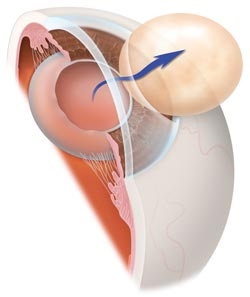
- The procedure may be followed by the implantation of an intraocular lens (L.O.L). The lens prosthesis is selected by the surgeon prior to the surgery, and may either be purchased by “consignment” with a company, or kept in stock in the operating suite.
Position
- Supine

Instrumentation
- Basic eye procedure tray
- Cataract extraction tray
- Phacoemulsification tray
- Intraocular lens implant
Supplies/ Equipment
- Basin set
- Balanced saline solution
- Irrigation/ aspiration pack
- Ophthalmic sponges
- Ophthalmic cautery
- Microscope drape
- Headrest
- Sitting stool with backrest
- Cryoextractor
- Phacoemulsifier
- Honan intraocular pressure reducer cuff
- Beaver blade
- Super blade
- Multipore filter
- Medications
- Sutures
Procedure
Intracapsular
- A lid speculum is placed and traction sutures are placed in the sclera.
- The conjunctiva is reflected from the superior cornea.
- Bleeders are cauterized.
- The anterior chamber is entered; an iridotomy is performed as the cornea is retracted by suture traction.
- An enzymatic solutioin is instilled into the anterior chamber to dissolve the zonule fibers suspending the lens.
- A cryoextractor is applied to the lens, which adheres to it, and the lens is withdrawn from the eye.
- The corneal incision is closed; traction sutures are removed, and the conjunctival flap is approximated.
- If an intraocular lens implant is used, it will be implanted following the extraction of the lens.
- The prosthesis is either sutured to the iris or simply held in place by the iris, depending on the type of prosthesis.
- Ophthalmic ointment may be instilled, and an eye dressing and patch is applied.
Extracapsular
- This procedure is similar to the intracapsular procedure, except that the lens capsule is incised, and the lens is exposed or irrigated out leaving the posterior capsule, which remains as a barrier to the vitreous humor.
- When phacoemulsification is used, the anterior lens capsule is excised.
- The lens nucleus is prolapsed into the anterior chamber, and the ultrasonic probe is inserted into the capsule.
- The probe is set to irrigate/ aspirate and then fragment the remaining lens substance.
- After the “phaco” procedure, the wound is closed.
Perioperative Nursing Considerations
- If a floor model microscope is used, it should be draped and brought in over the field on the opposite side of the affected eye.
- Thorough familiarity with all equipment used its mandatory for a smooth surgical procedure
- Check all the equipment before use.
- The circulator will usually be responsible for changing the settings on the phacoemulsifier unit.